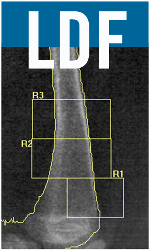
Distal Femur Analysis – Lunar Prodigy Advanced
Scan modality used: Forearm
Analysis Instructions:
1) Select scan to be analyzed
Determine where bone is:
View the image to be sure that the bone edges are correct and that it filled properly.
2) If necessary, click Points button.
3) Fill in/clean off bone edges as necessary.
4) Click Results button.
Draw regions of interest:
5) Select Analyze pull down screen.
6) Click Custom.
7) Click ROI’s button.
8) Along the left hand side of the screen, click on the rectangular box (create rectangle).
9) Click on box that allows you to move the entire rectangle
10) Move the box to the top of the femur where the shaft is a constant width, placing the upper left corner
on the left bone edge.
11) Click on the right vertical side of the rectangle and drag it so the upper corner is on the right bone edge.
12) Make notation of the width of this measurement
13) Refer to the chart – look at left side (Width of Top of Femur) to determine the height of R1
or (Width at the top of the femur x 0.5714)x2 = Height of the R1
14) Make height of the box this number.
15) Grab the rectangle and move it to the growth plate. The bottom of the box should sit just above the
space at the growth plate. If the growth plate is angled, place the bottom of the box in the middle of
the growth plate. (If the growth plate is angled you will need to “tip” the box. Use the move vertex
button on the left side of the screen, being sure to count the number of times you move the vertical
line to gain the correct angle. You will need to tip both vertical lines the same amount to ensure you
maintain a parallelogram.)
16) Determine width of femur at the growth plate.
17) Refer to right side of chart (Width of Base of Femur)
18) Move back of the ROI box to this number (the center of the femur)
or
half of the width of the femur at the growth plate
19) Move the front of the ROI box into the space in front of the femur (beyond the bone edge).
20) This is R1.
Duplicate Regions:
21) Click on the rectangular box (create rectangle).
22) Create another rectangle on top of R1 making it the same height but a different width.
23) Click on Move/size ROI button.
24) Move the new copy so that it lies directly above R1.
25) Click on the vertical sides to extend both horizontal edges of the new box beyond the bone edges so
there is an even amount of space on each side of the femoral shaft.
26) Click on the rectangular box (create rectangle).
27) Click on Move/size ROI button.
28) Move the new copy so that it lies directly above R2.
29) Click on the vertical sides to extend both horizontal edges of the new box beyond the bone edges so
there is an even amount of space on each side of the femoral shaft.
Results:
30) Click Results.
The aspect ratio of your machine is the correction factor used in determining the height of the ROI boxes
Correction Factor = Pixel Width/Pixel Height
Lunar Prodigy Advanced:
Forearm Pixel Width= 0.6 mm
Forearm Pixel Height = 1.05 mm
Distal Femur Analysis – HOLOGIC (Fan Beam Machines)
Set up for analysis:
1) Select Analyze Scan.
2) Select Unanalyzed Scans.
3) Double-click on the patient’s scan.
4) Select Subregion Forearm as the Analysis Method.
5) Click on Next.
Determine where bone is:
1) Click on bone map box.
2) If necessary, click on Add Bone or Delete Bone. Use the mouse to draw the bone edge and/or add
bone. Sink Islands will automatically delete the areas outside the line. Fill Holes will automatically fill in bone.
Draw regions of interest:
1) Click on Subregions.
2) Click on “+” key to create an ROI box
3) Click on Whole Mode
4) Move the box to the top of the femur to a point where the femoral shaft is a constant width.
5) Click on Line Mode.
6) Determine the width of the femur (outside edges of cortical bone). Move the vertical edges of the box
to the edges of bone.
7) Refer to chart* – look at left side (Width of Top of Femur) to determine the height of R1
Or
(width at the top of the femur x 0.4226*)x 2 = height of the R1
8) Make height of box this number.
9) Click on Whole Mode and move the ROI box to the growth plate. It should sit just above the growth plate.
If the growth plate is angled, place the bottom of the box in the middle of the growth plate.
10) Click on Line Mode.
11) Determine width of femur at the growth plate.
12) Refer to right side of chart (Width of Base of Femur)
13) Move back of the ROI box to this number (the center of the femur)
Or
half of the width of the femur at the growth plate
14) Move the front of the ROI box into the space in front of the femur (beyond the bone edge).
15) This is R1.
16) Click on Whole Mode.
17) Click on the “+” key to copy the R1 box.
18) Move the new copy so that it lies directly above R1.
19) Click on Line Mode and extend both horizontal edges of the new box beyond the bone edges
so there is an even amount of space on each side of the femoral shaft.
20) Click on Whole Mode and click on “+” key to make additional copies of the ROI boxes.
21) Make two subsequent ROI boxes moving up the bone until there are a total of three ROI’s. ROI boxes
may be stair-stepped if necessary due to an angled femur.
Results:
1) Click on Results.
2) Click on Close
Add z-scores to DXA Report:
1) Open z-score calculator and calculate z-scores
2) Make note of numbers by ROI
3) Click on Report
4) Click on DICOM/IVA Report
5) Click on Scan Details
6) Under “Scan Comment”, type: Z-Scores: R1 = <enter the R1 z-score>; R2 = <enter the R2 z-score>;
R3 = <enter the R3 z-score>.
7) Click OK
Sending through PACS: Only if your facility has PACS and DXA is linked to PACS
1) Click on Send
Printing a Report: If you require a paper copy
1) Select Report
2) Check Filing
3) Click on Print
* The aspect ratio of your machine is the correction factor used in determining the height of the ROI boxes
Correction Factor = Pixel Width/Pixel Height


